Eye exams involve a series of tests that are done to evaluate your vision and check your eyes for any diseases or complications.
Eye examinations require the use of a variety of instruments and procedures for evaluating the eyes.
In this article, we’ll focus on some of the more common and medically important eye tests. If you found any problem in your eye vision you can treat it from medication too, TheyaVue Buy Online.
Each of these tests evaluates several aspects of your eye and can be useful in determining a variety of diseases, i.e. ophthalmological examination.
Signal
Most eye tests are indicated if you are having problems with your eyes or your vision (seeing things). Some common indications for an eye exam are:
- Severe Eye Injury
- Eye Pain
- Intense Red Eye
- Sudden Vision Loss
- Eye Discharge
- Double Vision (Diplopia)
- Blurred Vision
- The Light Shines
- Chronic diseases like diabetes, multiple sclerosis
As A General Check
Tests are done at different ages throughout your life by your doctor to make sure your eyes are working properly. These routine check-ups are done for:
Child – Your pediatrician will examine your child’s eyes for healthy eye development and look for any problems like lazy eyes or cross-eye etc.
For children ages 3 to 5 your doctor may examine your child’s eyes for vision and eye development.
Adult- It is recommended that you get an eye exam around the age of 40, regardless of whether there is a problem or not.
This is the age when some common eye diseases begin to appear and cause problems, so they can be treated at the earliest.
If you are 60 years of age or older, you are recommended to have an eye exam every year or two. If you wear glasses or contact lenses, have a family history of eye problems, or take certain medicines that are toxic to the eyes.
Risk
Normal eye examination is superficial (non-penetrating) and does not cause any damage to the eyes or any body structure.
Most detailed eye exams require the use of some pupil dilating eye drops. These eye drops usually do no harm and you will just need to wear your sunglasses after your exam.
However, some adverse effects that may be caused by diluting eye drops include:
- Attack Of Narrow-Angle Glaucoma (Especially If You Are Older)
- Dizzy
- Flushing And Dry Mouth
- Nausea Or Vomiting
During some procedures, your doctor may use some anesthetic drops to numb your eyes. This local anesthetic may make you feel numb around your eye after your exam.
Patient Preparation
An ophthalmologist will do a thorough examination of your eyes, IE your visual acuity or diagnose complex diseases, and also perform surgery if needed.
You should consider taking the following precautions:
- If you wear glasses or contact lenses, you should bring them with you so that your doctor can make sure they are still the best fit for you.
- You should bring sunglasses with you to wear them after your exam, as some procedures can make your eyes sensitive to sunlight.
- You should also consider that someone else is driving you back home.
Refraction Assessment
This evaluation is used to determine the best refractive power for your contact lenses or glasses. Your doctor will determine the strength that will help you have the sharpest vision.
Retinal Probe
This is the main examination performed by an ophthalmologist and is also called ophthalmoscopy or fundoscopy.
Your doctor does a thorough evaluation of your eyes, including your retina (the back of your eye), the optic disc, and the retina’s blood vessels.
Your doctor will use eye drops to dilate your pupils. The pupils allow the amount of light to enter your eyes. The dilated pupil allows your doctor to see the inside of your eye in detail.
You will be asked to lie on a flat or against a chair.
Eye drops used prior to the procedure can sometimes cause some adverse reactions as described earlier.
Fundoscopy can be done in two ways. After administering the drops your doctor may use:
Direct Approach – Your doctor will shine a beam of light directly through your pupil to look at the back of your eye. Both your eyes will be examined.
Indirect Method – This is a very useful method of assessing your retina as it gives a three-dimensional view of the structures present in your eye.
Your doctor will use a condensing lens and a bright light placed on their forehead.
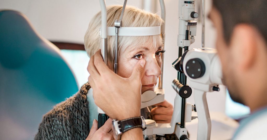
Lamp Ophthalmoscopy – you will be sitting in a chair with your chin on the device placed in front of you and your forehead in place so that you do not move.
Your doctor will then use the microscope part of the slit-lamp and some other small lens to visualize your retina. This procedure allows for even greater magnification of the internal structures of the eye.
Ophthalmoscopy can be used to detect certain abnormalities within your eye, especially those related to the retina. Some problems can be detected by this process:
Retinal inflammation, such as CMV retinitis, diabetic retinopathy
- Eye Disease
- Acute Age-Related Macular Degeneration, Leading To Loss Of Central Vision
- Eye Melanoma
- Optic Nerve Problems
- Any Problem Related To Blood Vessels
- Retinal Detachment